
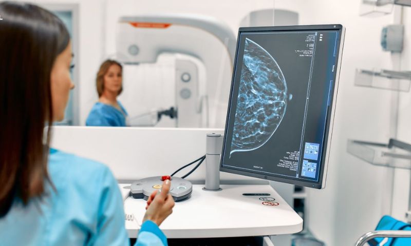


NIJMEGEN, Netherlands: Radiologists using artificial intelligence (AI) in reading mammograms were able to concentrate more on suspicious regions, according to a recent study published in a radiology journal.
A mammogram is an X‑ray image of the breast used to detect abnormalities, and when breast cancer is identified early through routine screening, it offers the best chance of effective treatment.
While earlier research showed that AI enhances cancer detection accuracy, until now, the effect on radiologists’ visual search patterns was unclear. In this study, eye‑tracking technology was employed to compare how radiologists reviewed screening mammograms with and without AI assistance.
The results demonstrated that, with AI support, radiologists gave significantly more time to examining regions flagged as suspicious, serving as a valuable guide to where attention was most needed.
This study highlights that AI not only improves detection but also subtly alters radiologists’ workflow, enabling them to focus more precisely on potential abnormalities.
“By analysing this data, we can determine which parts of the mammograms the radiologist focuses on, and for how long, providing valuable insights into their reading patterns,” says joint first author Jessie Gommers, from the Department of Medical Imaging, Radboud University Medical Center in Nijmegen, Netherlands.
A small eye-tracking device, featuring two infrared lights and a central camera, was positioned in front of the mammogram display. The infrared lights illuminated the radiologist’s eyes, and the camera recorded reflections to precisely map their gaze coordinates on the screen.
Analysis of the eye-tracking data revealed that when aided by AI, radiologists spent notably more time examining areas containing actual lesions, without any increase in overall reading time. This AI assistance also translated into improved breast-cancer detection accuracy across the board.
“The results are encouraging,” Gommers says. “With the availability of the AI information, the radiologists performed significantly better.”
“Radiologists seemed to adjust their reading behaviour based on the AI’s level of suspicion: when the AI gave a low score, it likely reassured radiologists, helping them move more quickly through clearly normal cases,” Gommers says.
The study involved 12 radiologists who read mammography examinations for 150 women, 75 with breast cancer and 75 without.
“Educating radiologists on how to critically interpret the AI information is key,” says Gommers.
Overall, AI not only helped radiologists focus on the right cases but also directed their attention to the most relevant regions within those cases, suggesting a meaningful role for AI in improving both performance and efficiency in breast cancer screening.